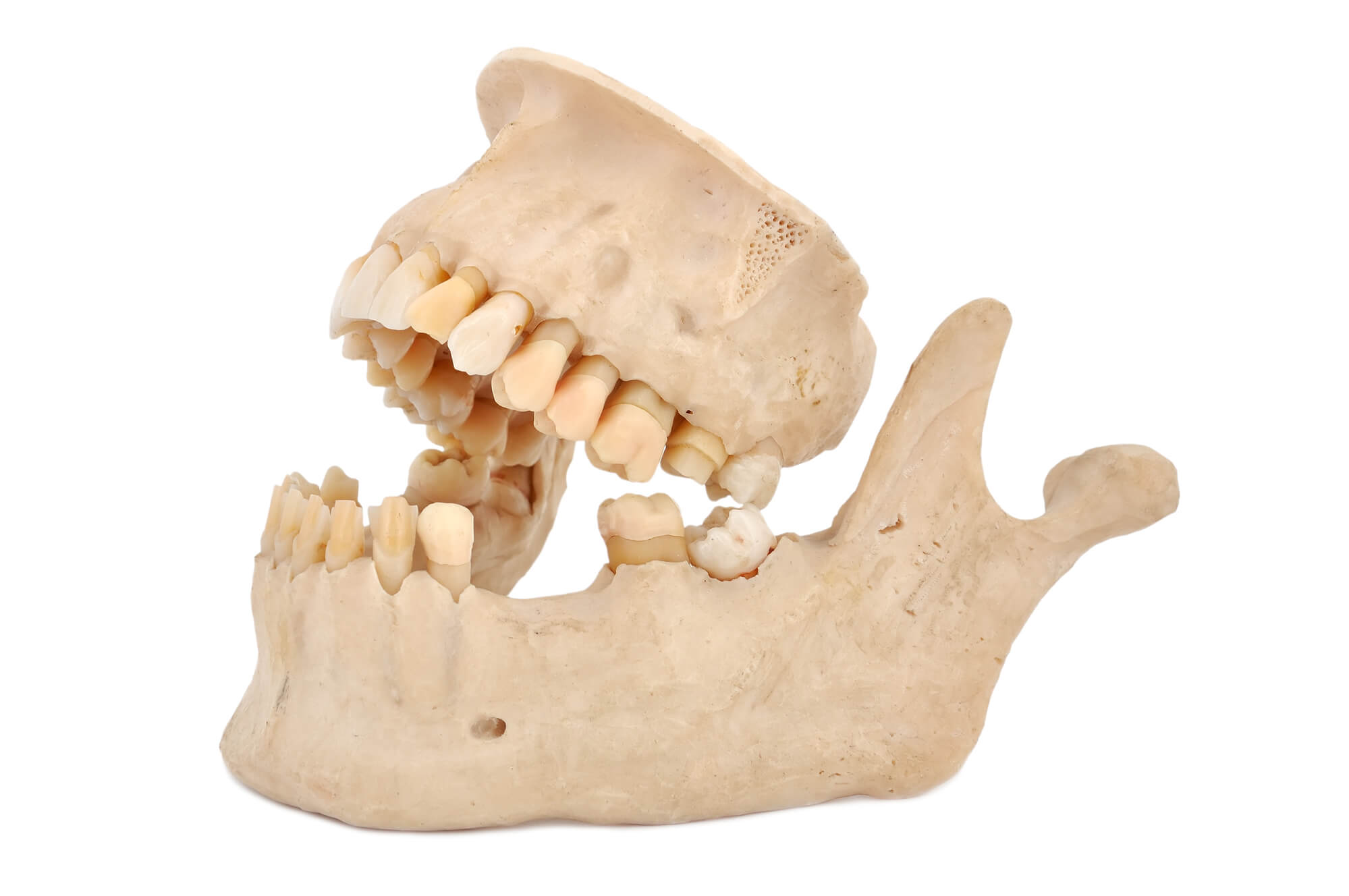
Osteo-cavitations, or areas of dead bone are also called NICO lesions. This stands for neuralgia -inducing cavitational osteonecrosis. They are hollow places in your jawbones. Interestingly, the concept of jaw bone cavitations is very controversial. Many in the dental profession refuse to believe that these bony lesions even exist. However, in the specialties of orthopedics, neurosurgery and internal medicine such lesions are well recognized.
Many of these lesions are quite painless, lying dormant, like a smoldering fire, often for decades. At least 20% of all hip replacements in the USA are due to osteonecrosis. Why many in the dental profession deny the existence of NICO lesions, remains a mystery.
These lesions have been defined as a cavity within a cavity. I call them, just another hole in your head that you don’t know about.
The mandible and the maxilla, like the hip, are medullary bones. This means they have bone marrow and they produce blood cells. It is usually within these medullary spaces, that cavitations occur. Blockages in tiny blood vessels reduce blood flow, thus robbing oxygen from areas of the bone, causing bone death. This process is similar to a stroke.

The diagnosis of cavitations can be hindered by the fact that x-ray examination is often times unremarkable. Considerable diagnostic experience is required because changes in the bone are subtle and may mimic normal bony anatomy. Osteonecrosis is a disease of the marrow spaces of the bone and 30 to 50% of the bone must be destroyed before changes can be seen on your x-rays.
Better Diagnoses Using MRI Imaging Scans
MRI imaging scans work very well for long bones, but the flat bones of the face are not imaged well with normal MRI scans. The new CBCT scans which are three-dimensional are more effective in determining cavitations. Cavitat bone scanning ultrasounds are somewhat effective and accurate, especially when compared with patient’s panoramic x-rays. Surgical success is improved when using these imaging techniques.
You must have your dental surgeon anesthetize the area, in order to determine whether a cavitation exists or not. If the pain goes away after an anesthetic injection, then you can be reasonably certain there’s a problem in that area generating the pain.
A new ultrasound image technique called, MRI - STIR imaging, has shown remarkable accuracy in determining cavitation locations.
There have been over 74 risk factors isolated and identified which contribute to cavitation formation. These factors apply to all bones, including the dental arches. Trauma and infection are the primary triggering causes for cavitation’s. There are no other bones in the body that come close to the amount of trauma and infection that occur in the maxilla and the mandible.
Cavitations may be completely painless. Chronic inflammation and swelling can occur in one or more of the tiny vessels producing the death of the vessels, then ischemia occurs, and ultimately bone death.
Causes of Cavitations
- Trauma, maybe from mild or severe direct blows to the jaw
- Persistent tooth infections, chronically sensitive teeth after dental restorations or severe tooth infections
- Surgical trauma, following surgical extractions, endodontic surgeries etc.
- Thrombophilia and hypo-fibrinolysis, these are genetic blood coagulation disorders that predispose some people to form blood clots thereby preventing blood flow, which starves the bone of oxygen. The first condition increases the tendency to develop blood clots. The second condition reduces the ability to destroy blood clots.
- Failed root canals, chronic persistent infections following root canal therapy
- Anti-phospho-lipid syndrome, with this immune system disease, blood clots can develop within small artery and veins. This is an autoimmune response. With APS, antibodies are manufactured and act against your body, often producing blood clots and cavitations if the clots occur within the bone.
- Extractions, usually third molar extractions, but can be any tooth
Cavitation Treatment
Here are a few modalities of therapy used to treat osteocavitations:
- Surgery with PRP, surgical debridement of the affected area and the introduction of protein rich plasma, called PRP, which facilitates growth of new bone. All materials removed during the surgery must be biopsied. This process removes the dead bone and bone marrow, and creates a new environment for healing.
- Hyperbaric therapy, this is similar to ozone therapy, but it is a systemic treatment technique. You are placed in a hyperbaric chamber, which raises the atmospheric pressure and drives oxygen into your cells.
- Anticoagulant therapy, if you have a systemic clotting problem or other clotting risk factors, specific anticlotting medications can be used. Check the list in module 4, which contains natural anti-blood clotting therapies.
- Injection of homeopathic remedies, this has become very popular recently, but is not a permanent solution because the dead bone has no blood supply, which is required for normal healing and removal of toxins
- Ozone therapy, injections of ozone directly into the cavitation site. This is a very effective treatment modality.
- Natural antibiotic therapies, see the list in the biological dentistry module.
What about Surgical Failures?
- Osteonecrosis has a strong tendency to recur or redevelop additional lesions.
- This happens about 30 to 40% of the time.
- 5 to 10% of the time little or no reduction in pain occurs.
- About 15% of the time moderate reduction in pain occurs.
- Often and sadly, nearly 3% have an increase in pain after surgery.
- Make sure any and all material is removed from the cavitation site during surgery, then biopsy the site, including the teeth.
Common Cavitation Symptoms
- Deep bone pain, which is constant, but varies in intensity.
- Sharp shooting pain, which is hard to diagnose.
- Chronic maxillary sinusitis including congestion and pain.
- A sour bitter taste, which can cause gagging and bad breath.
- History of large fillings, followed by pain, then root canals, then removal of the tooth.
- Multiple rooted canals
- Endodontic apicoectomy
- Tooth extraction
- Failed trigeminal neuralgia treatment
Most common scenario’s start with a simple dental restoration. Many times NICO lesions produce no symptoms at all, however, these lesions can also produce intense, trigeminal neuralgia-like symptoms.
There are established characteristic referred pain patterns, usually with an underlying constant dull ache. In addition to the deep ache, there can be a sharp shooting pain in the area of the trigeminal nerve. If you suspect a cavitational problem, allow no one to operate without first proving the pain can be stopped with a local anesthetic injection. This is very important.

If the pain can’t be stopped for a short period of time with a local anesthetic injection, it’s a good bet that another problem is occurring along with the cavitation. Make sure you request a cavity ultrasound examination. Ask your surgeon what his or her success rate is. If you are told more than 90% get another opinion, find another doctor, or turn and run.
What about Post-Operative Complications?
Due to the anatomical complexities of the upper and lower jaws, you must weigh the complications against the benefits of surgery.
- Pain
- Bruising
- Infection or swelling
- Partial improvement
- No improvement
- Worsening of the condition
- Damage to nerves
- Entrance into the maxillary sinus
- Injuries to the TMJ
- Loss of one or more teeth
- The need to have the surgical procedure repeated
- Mutilation or damage to the bony areas
Fortunately, these complications occur only rarely, but if you’re considering cavitation surgery you must know these possible complications and ramifications. If your doctor doesn’t know about these complications or doesn’t want to discuss them, find a new doctor.
Source: TMJ and Facial Pain Center, Minneapolis, Minnesota
Life Rejuvenation Systems | All Rights Reserved | www.DrLokensgard.com


